
FIGURE 2.
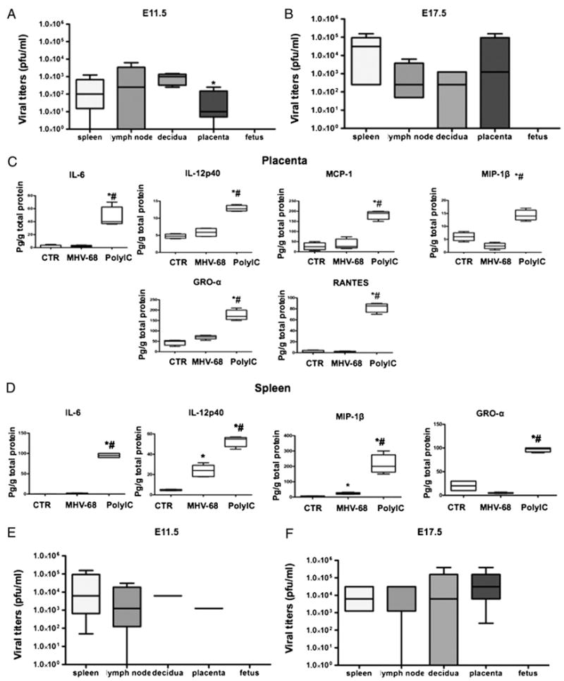
Effect of MHV-68 viral infection in pregnant mice. Viral titers as PFU/ml were determined in wt pregnant mice infected with MHV-68 (1 × 10⁶ PFU) 3 d (E11.5; A) and 9 d (E17.5; B) postinfection. Viral titers were observed in lymph nodes, placenta, decidua, and spleen, but were absent in the fetuses. *p < 0.05, decidua versus spleen. Placenta (C) and spleen (D) cytokine profile was determined in wt pregnant mice treated with poly(I:C) or MHV-68 4 and 9 d postinfection, respectively. *p < 0.05, MHV-68 versus control; #p < 0.05, poly(I:C) versus MHV-68. E and F, Viral titers as PFU/ml were determined in TLR-3 KO pregnant mice infected with MHV-68: E, 3 d (E11.5), and F, 9 d (E17.5) postinfection. Note the high levels of viral titers in lymph nodes, placenta, decidua, and spleen, but absent in the fetuses. Bars show median ± SEM. n = 6 mice per group.