Figure 2.
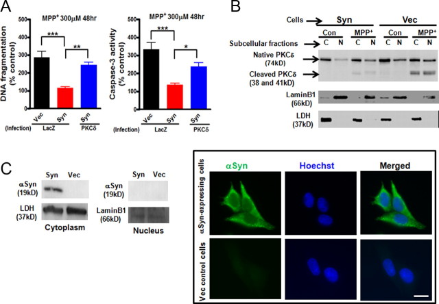
Deregulation of PKCδ by α-synuclein protects against MPP+-induced cell death in dopaminergic N27 cells. A, Effects of downregulation of PKCδ by αsyn on MPP+-induced cell death in dopaminergic N27 cells. αSyn-expressing (Syn) and vector control (Vec) N27 cells were infected with lentiviruses expressing LacZ-V5 or PKCδ-V5 for 24 h. The cells were then exposed to MPP+ (300 μm) for 48 h. Cells were collected and assayed for DNA fragmentation (left panel) and caspase-3 activity (right panel). Data shown represent mean ± SEM from two independent experiments performed in quadruplicate (*p < 0.05; **p < 0.01; ***p < 0.001). B, MPP+-induced PKCδ proteolytic cleavage and its nuclear translocation were significantly diminished in αsyn-expressing N27 cells. αSyn-expressing (Syn) and vector control (Vec) N27 cells were exposed to MPP+ (300 μm) for 36 h. Cytoplasmic (C) and nuclear (N) fractions were prepared for immunoblotting analysis of PKCδ. LDH (cytoplasmic fraction) and Lamin B1 (nuclear fraction) were used as loading controls. C, Cytoplasmic localization of αsyn in αsyn-expressing N27 cells was not affected by MPP+ treatment. αSyn-expressing (Syn) and vector control (Vec) N27 cells were exposed to MPP+ (300 μm) for 36 h. Cells were either collected for preparation of cytoplasmic and nuclear extracts and immunoblotting analysis of αsyn (left panel) or stained and visualized under a Nikon TE2000 fluorescence microscope (right panel). Scale bar, 10 μm. A representative immunoblot and image of αsyn immunostaining (green) and Hoechst staining (blue) are shown.

