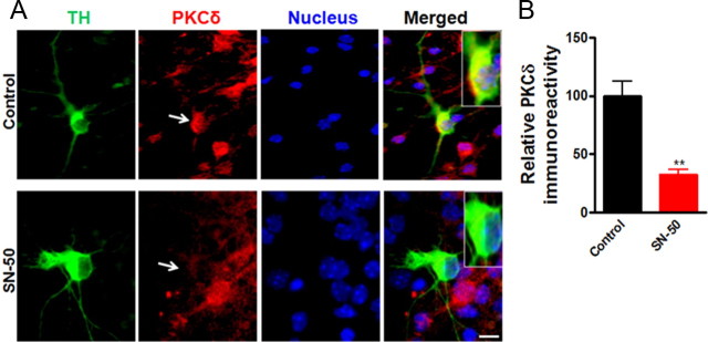
Figure 6.
Effect of NFκB inhibition on the PKCδ immunoreactivity in the primary dopaminergic neurons. A, Primary midbrain cultures were treated with or without 100 μg/ml SN-50 for 24 h. Cultures were immunostained for TH (green) and PKCδ (red). The nuclei were counterstained by Hoechst 33342 (blue). Images were obtained using a Nikon TE2000 fluorescence microscope. Magnification, 60×. Scale bar, 10 μm. Representative immunofluorescence images are shown. The inset shows a higher magnification of the cell body area. B, Immunofluorescence quantification of PKCδ in TH-positive neurons. Fluorescence immunoreactivity of PKCδ was measured from TH-neurons in each group using MetaMorph software. Values expressed as percentage of control group are mean ± SEM and representative for results obtained from three separate experiments in triplicate (**p < 0.01).

