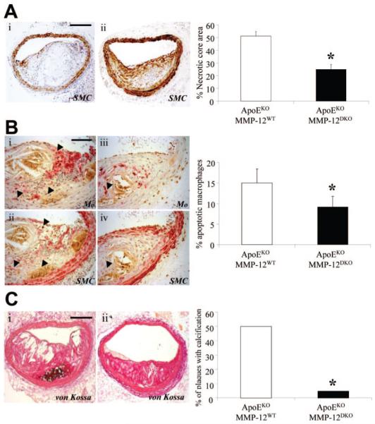
Figure 6.
Effect of MMP-12 gene deletion on apoptosis and calcification. A, Necrotic core area of brachiocephalic artery lesions from apoEKO/MMP-12WT and apoE/MMP-12DKO mice. SMC indicates smooth muscle cell. B, Immunohistochemical labeling of brachiocephalic artery lesions for macrophages, smooth muscle cells (both red) and apoptosis (brown, arrows). C, Presence of calcification (brown/black, arrows) in brachiocephalic artery lesions from apoEKO/MMP-12WT and apoE/MMP-12DKO mice. Quantification is summarized in the adjoining graphs and depicts mean±SEM (*P<0.05, n≥23 per group). Scale bars represent 100 μm and are applicable to adjoining panels.

