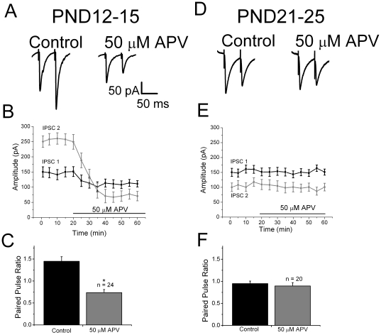
Figure 1. Physiological evidence for modulation of GABAergic IPSCs by presynaptic NMDA receptors in rat neocortex.
A: in slices from PND12-15 animals, paired pulse facilitation was observed under control conditions (left trace). After bath application of D-APV, the same stimulation evoked a smaller IPSC 1 and subsequent paired pulse depression. These results suggest the presence of tonically activated facilitatory presynaptic NMDA receptors. B: time course plot of IPSC 1 and IPSC 2 amplitudes showing effect of D-APV on each response. C: summary plots show the effect of D-APV on paired pulse ratios in PND12-25 group. D: specimen records from PND21-25 animals showing paired pulse depression under control conditions. Responses did not change after wash in of APV. E: time course plot of IPSC 1 and IPSC 2 amplitudes showing that D-APV had no effect on either response in the PND21-25 group. F: summary plots show developmental changes in the effect of APV on paired pulse ratios. IPSCs were pharmacologically isolated by recording in the presence of 50 µM GYKI52466, 20 µM CNQX and 2 µM SCH50911 to block AMPA, KA and GABAB receptors, respectively. The recording pipette contained 1 mM MK-801 to block postsynaptic NMDA receptors. Specimen records shown are averages of 10 responses.

