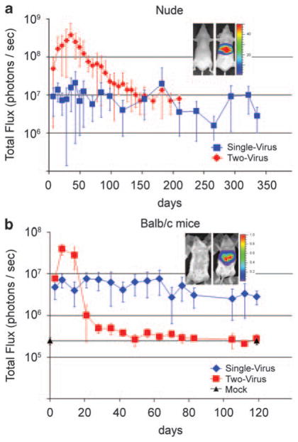
Figure 3.
Persistence of transgene expression in vivo assayed by bioluminescence imaging of luciferase activity in the liver of the same mice over time. (a, red) A single tail vein injection of 3.2 × 10⁸ IGU of the single-virus vector HDAd-EBV.1x.hRluc (Figure 1a) was administered to nine nude mice. Of these, one died at day 98, two at day 154 and two at day 238, from causes unrelated to vector administration; four mice remained at the final time point of 336 days post-vector administration. At each time point indicated, all remaining mice were assayed for luciferase expression in the liver by bioluminescence following tail vein administration of the Renilla luciferase substrate coelenterazine, as described.14 (Inset shows bioluminescence images of a control (left) and a transduced (right) animal. Color scale 1, 50 × 106 photons per sec.) (blue) A second group of 16 mice were administered 5 × 10⁹ each of the two HDAds of the two-vector system (Figure 1b). Mice (number at each time point given in Supplementary Table 1) were assayed for bioluminescence from the region of the liver at the indicated times after injection. (b, red) Eight immunocompetent Balb/c mice were administered a single tail vein injection of 7.2 × 10⁸ IGU of the single-virus vector HDAd-EBV.1x.hRluc, or (blue) six Balb/c mice were administered 7.2 × 10⁸ IGU each of the two-virus vector system. Luciferase activity in the region of the liver was quantitated by bioluminescence at the indicated days post-vector administration.

