
Figure 1.
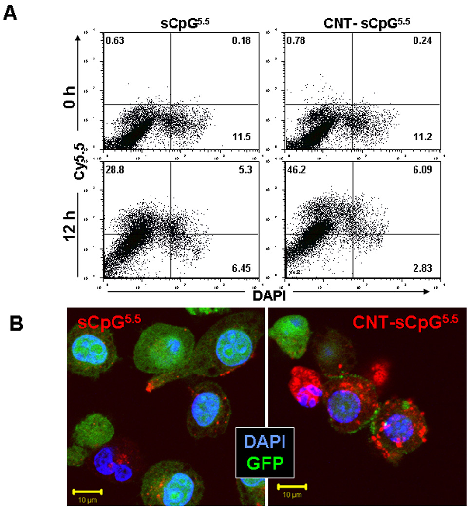
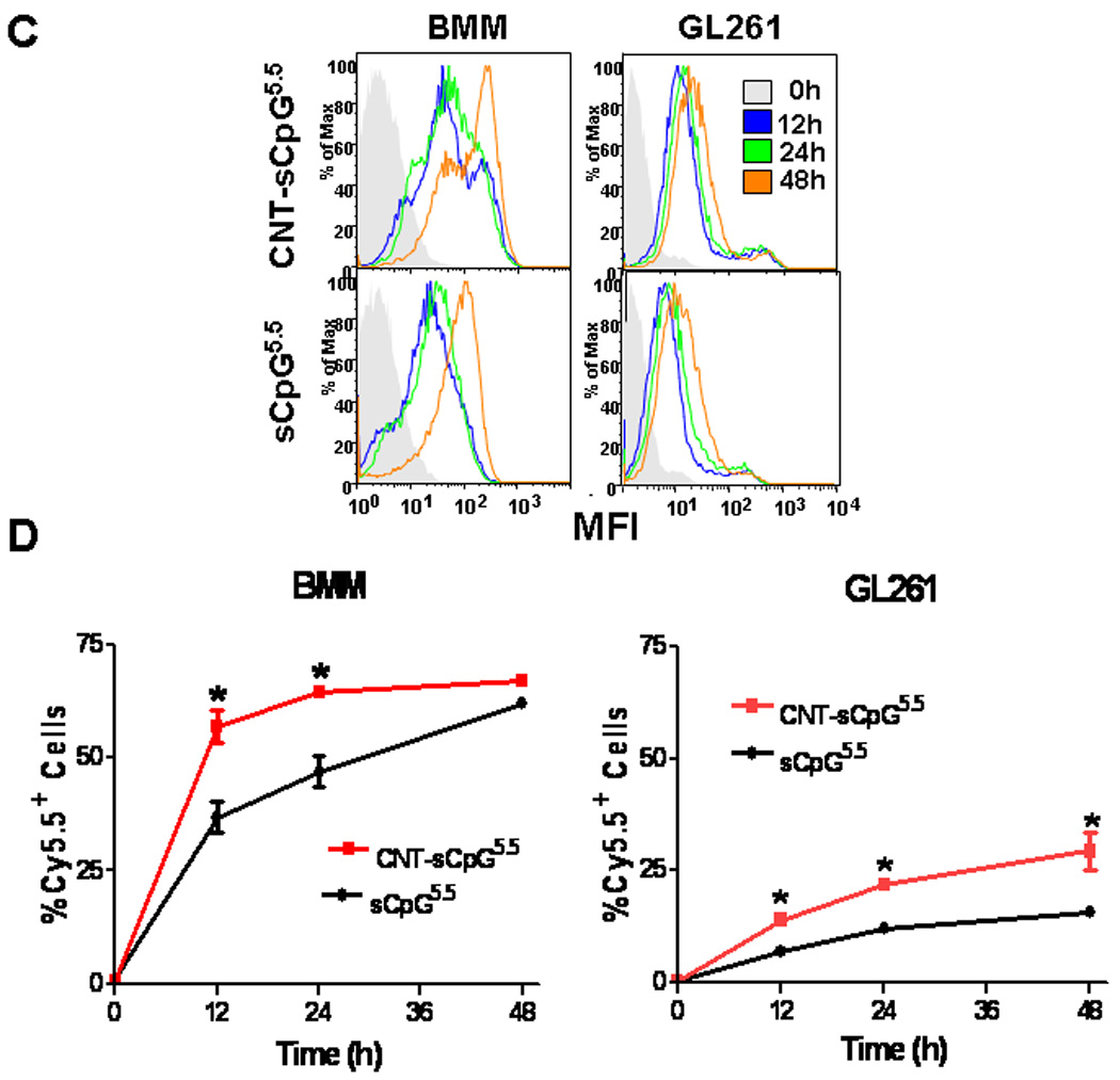
CNTs enhance CpG uptake in vitro. Primary bone marrow-derived monocytes (BMM) from wt or CX3CR1GFP mice or GL261 gliomas were incubated with PEG-functionalized single-walled CNTs conjugated to Cy5.5-labeled sCpG (CNT-sCpG5.5, 2.5 µg CNT- 5µg sCpG5.5/ml) or free sCpG5.5 (5µg/ml) and sCpG5.5 uptake was visualized by fluorescent microscopy and quantified by flow cytometry. A, dot plot demonstrating CNT-sCpG uptake by BMM to be more efficient than free sCpG5.5. B, CpG5.5 signal was stronger in BMM.gfp cells incubated with CNT-sCpG and was mostly confined to cytoplasmic compartments. C, histogram and D, time course experiments demonstrating CNT’s to also enhance sCpG5.5 uptake by GL261 gliomas, but not as efficiently as in BMM. Data is representative of three separate experiments. (n=3 samples /point, ±SEM); *, P<0.05, t-test.