Fig. 1.

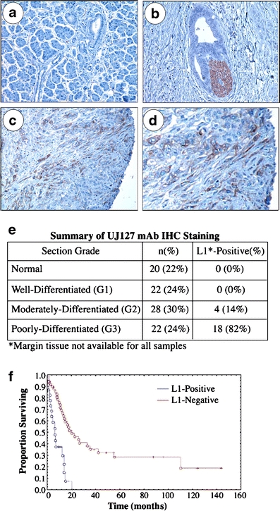
L1 is expressed by poorly differentiated PDAC cells in situ. UJ127 mAb visualized with DAB chromogen (brown). a Normal pancreas. b A well-differentiated tumor duct invading perineurally. Nerve bundle, brown. c Poorly-differentiated PDAC tumor cells at the margin. d High power image of c. e Immunohistochemical summary. f Kaplan–Meier analysis of patient survival
