
Fig. 1.
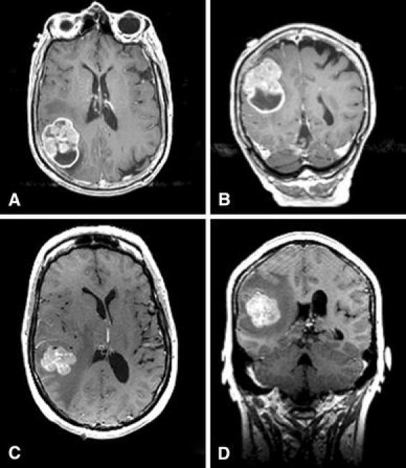
T1-weighted gadolinium-enhanced pre-operative MRI of the brain from Case 1 in axial a and coronal b sections demonstrating an irregularly enhancing and partially cystic lesion in the right parietal lobe, with minimal surrounding edema. T1-weighted gadolinium-enhanced pre-operative MRI of the brain from Case 2 in axial c and coronal d sections demonstrating an irregularly enhancing lesion in the right posterior temporal lobe, with minimal surrounding edema and shift of the midline structures to the left