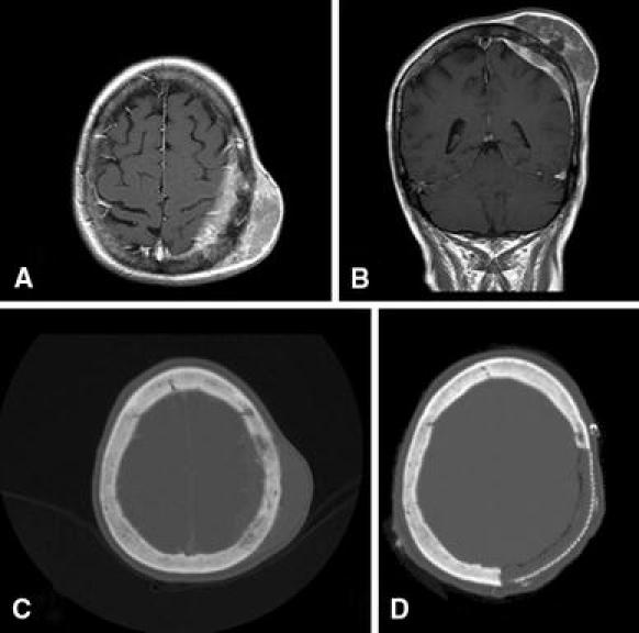
Fig. 2.

T1-weighted gadolinium-enhanced pre-operative MRI of the brain from Case 3 in axial a and coronal b sections demonstrating a subgaleal mass with involvement of the underlying bone and dura. Pre-operative axial head CT with bone windows c scan demonstrates subtle bony change with slightly irregular hyperostotic calvarium, but no gross osteolysis. Postoperative axial head CT shows the region of calvarial reconstruction with titanium mesh and methylmethacrylate d
