
Fig. 3.
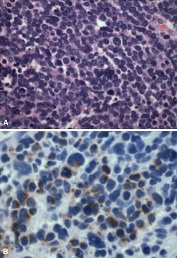
Case 2 a Light micrograph of the metastatic MCC specimen stained with hematoxylin and eosin at ×60 magnification shows a highly cellular specimen comprised of small to medium sized round cells with high nuclear to cytoplasmic ratio. The oval nuclei have a fine granular chromatin pattern. Mitotic figures were evident. b Light micrograph of the metastatic MCC specimen stained with CK-20 at ×60 magnification displays the paranuclear dot staining pattern typical of MCC