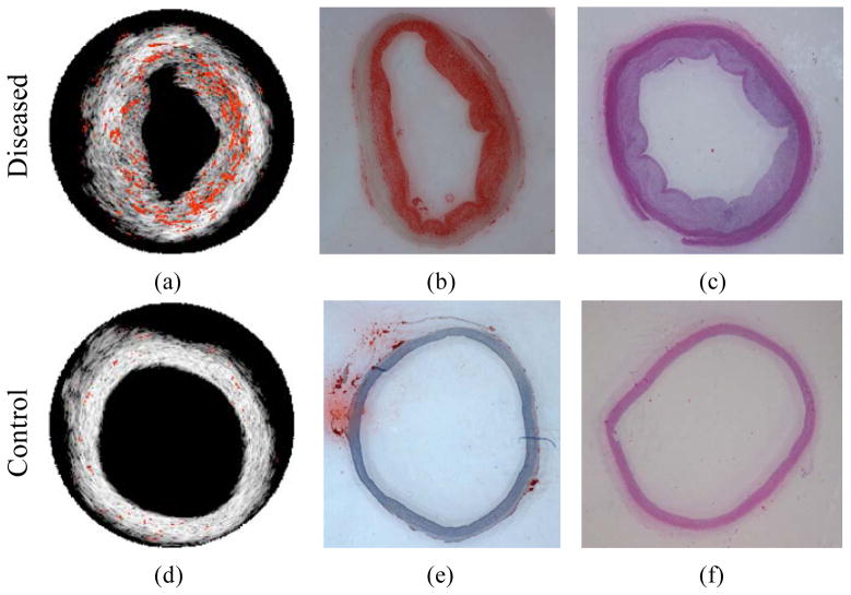
Figure 2.
Lipid regions (orange color) were demonstrated on top of the IVUS images for diseased (a) and control (d) rabbit aorta. (b, e) Oil Red O stain for lipid and (c, f) H&E stain closed to the imaged cross-section of diseased and control rabbit aortas. Reproduced with permission from Wang B, Su JL, Amirian J, et al. Detection of lipid in atherosclerotic vessels using ultrasound-guided spectroscopic intravascular photoacoustic imaging. Opt Express 2010 Mar 1;18(5):4889–97. [11].

