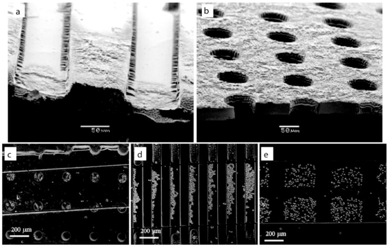
Figure 1.

PEG Hydrogel microstructures. Scanning electron micrographs of (a) PEG-bottomed microwells and (b) microwells exposed to the underlying substrate. Cell docking in various microstructures, including (c) round 100 μm diameter microwells, (d) grooves 50 and 75 μm in width (left and right respectively), and (e) 200 μm square microwells. Images reproduced with permission21.
