
Figure 4.
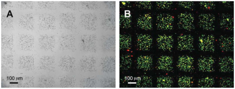
Light (A) and fluorescent (B) micrographs of micromolded cell-laden methacrylated hyaluronic acid (MeHA) hydrogels. The MeHA is crosslinked by exposure to light. The fluorescent micrograph shows cell viability after UV light exposure. Live cells are green (calecinAM) and dead cells are red (ethidium homodimer). Images reproduce with permission.33