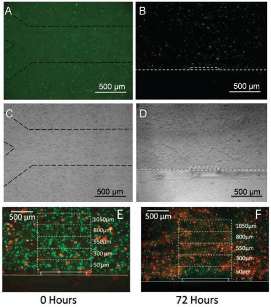
Figure 7.
Fluorescent and brightfield images of carboxyfluoresceinsuccinimide ester-stained AML-12 cells encapsulated in a microengineering agarose channel. (A, C) top view, and (B, D) are cross-sectional view of the channel. Dashed lines were added to images to aid in visualization at print resolutions.(E,F) Representative live/dead staining of AML-12 hepatocytes immediately after encapsulation and after 72 hours of culturing. Reproduced with permission.33

