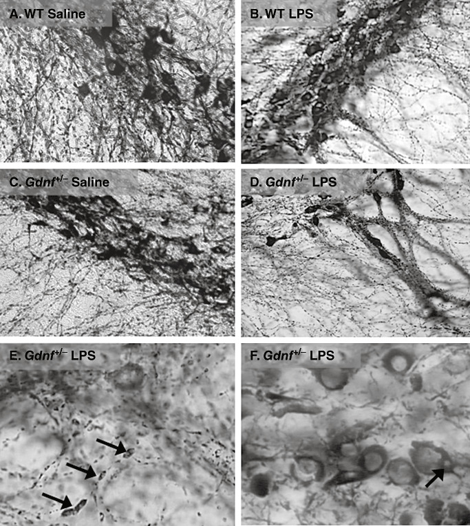
Figure 4.

Axonal swellings and cell body inclusions substantia nigra in lipopolysaccharide (LPS)‐treated mice. These close‐up images demonstrate a significant alteration in neurite and cell body morphology as a result of the prenatal LPS treatment in mice of both genotypes. The images are generated from 12‐month mice representing all groups (A–D), followed by two close‐up images (E,F) at high magnification showing the substantia nigra (SN) pc region of LPS‐treated Gdnf +/− mice at 12 months of age, with a high grade of axonal deterioration, pyknotic nuclei and cytoplasmic inclusions (see arrows in E and F, respectively). The tyrosine hydroxylase (TH)‐devoid inclusions were observed in many TH‐immunoreactive (TH‐ir) neurons at this age but only in mice treated with LPS. Abbreviation: WT = wild‐type.
