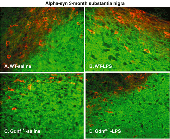
Figure 6.

α‐syn staining in substantia nigra (SN) at 3 months of age. Double labeling with α‐syn (green) and tyrosine hydroxylase (TH, red) of the SN region using fluorescence immunohistochemistry (A–D). Note that there were differences in the distribution of this protein between the pc (above) and the pr (below), in that both the Gdnf +/− saline and the wild‐type (WT) lipopolysaccharide (LPS) group had increased α‐syn staining in the pc, compared with the other two groups. At this age, few TH‐ir cell bodies (red staining) were double labeled with α‐syn (green), suggesting that α‐syn is expressed in other neuronal populations in this brain region as well.
