Figure 2.

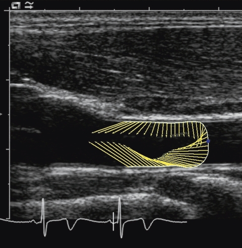
The image shows the common carotid artery (CCA) wall motion at the specific time point shown in the ECG recording. The vectors indicate the direction and magnitude of the CCA wall motion in the longitudinal and radial direction.

The image shows the common carotid artery (CCA) wall motion at the specific time point shown in the ECG recording. The vectors indicate the direction and magnitude of the CCA wall motion in the longitudinal and radial direction.