Figure 1.
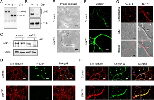
Establishment of JNK-deficient neurons. Wild-type (control) and Jnk1LoxP/LoxP Jnk2−/− Jnk3−/− CGNs were infected with Ad-cre at 3 d of culture in vitro (DIV) and then examined at 10 DIV. (A) Genotype analysis of JNKTKO neurons. The floxed Jnk1 and deleted Jnk1 alleles are detected as 1095-base-pair (bp) and 395-bp PCR products, respectively. (B) Extracts prepared from JNKTKO neurons were examined by immunoblot analysis using antibodies to JNK and α-Tubulin. (C) Control and JNKTKO neurons were examined at 10 DIV by immunoblot analysis using antibodies to phospho-neurofilament H and α-Tubulin. (D) Control and JNKTKO neurons were examined by immunofluorescence microscopy by staining with DAPI and antibodies to βIII tubulin and phospho-Ser63 cJun. Bar, 20 μm. (E) Control and JNKTKO neurons were examined by phase-contrast microscopy. Bar, 75 μm. (F) Control and JNKTKO neurons were stained with calcein-am ester and examined by fluorescence microscopy. Bar, 65 μm. (G) Wild-type (control) and JNKTKO neurons were stained with Mitotracker Red at 10 DIV and imaged by differential interference contrast (DIC) and fluorescence microscopy. Bar, 8 μm. (H) Control and JNKTKO neurons were examined by immunofluorescence microscopy by staining with DAPI and antibodies to βIII tubulin and Ankyrin G. Bar, 20 μm.

