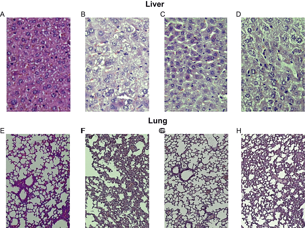
Figure 6.

Treatment with NDP-α-MSH (340 µg·kg−1 i.p. for 7 days after zymosan) reduced histological damage in the liver and lung of MODS mice. Representative light microscopy images of experiments of Table 1. (A) Normal liver histology at day 7 from sham MODS mice. (B) Representative liver from saline-treated MODS mice: presence of inflammatory cell infiltrates, steatosis, necrosis and ballooning degeneration. (C) Representative liver from NDP-α-MSH-treated MODS mice: few inflammatory cells, reduced steatosis, absence of necrosis and/or ballooning degeneration. (D) Representative liver from MODS mice treated with the nicotinic receptor antagonist chlorisondamine 1 min before each administration of NDP-α-MSH: massive necrosis, cellular degeneration, strong presence of inflammatory cells. (E) Normal lung histology at day 7 from sham MODS mice. (F) Representative lung from saline-treated MODS mice: presence of a sustained inflammatory cells infiltrate, vascular congestion and interstitial oedema. (G) Representative lung from NDP-α-MSH-treated MODS mice: reduction of inflammatory cells infiltrate, vascular congestion and interstitial oedema. (H) Representative lung from MODS mice treated with the nicotinic receptor antagonist chlorisondamine 1 min before each administration of NDP-α-MSH: strong inflammatory cells infiltrate, vascular congestion and interstitial oedema. Haematoxylin-Eosin staining; original magnification: liver X 40 and lung X 20.
