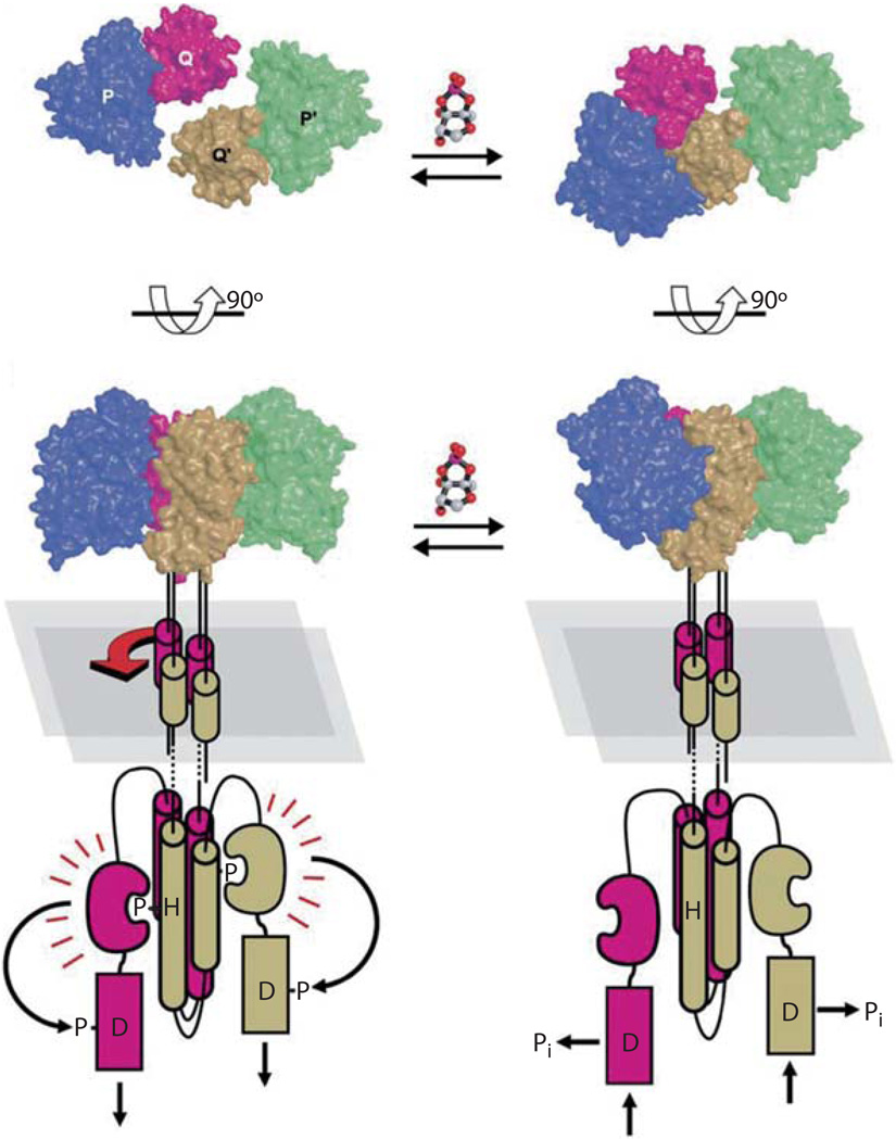
Fig. 4.
Model for AI-2 dependent LuxPQ receptor activity. The top panels display the LuxPQp receptor complex from a ‘top-down’ view, and the lower panels are a 90° rotation, showing the complex from the side. In the absence of AI-2 (left) LuxQ-LuxQ9 dimers exist in a symmetric orientation both in the periplasm and the cytoplasm, and therefore are in kinase mode. AI-2 binding to LuxP (right) causes a conformational change in LuxP, and induces interactions with LuxQ9. Simultaneous contacts of LuxP with LuxQ and LuxQ9 rotates the complex into an asymmetric orientation. Rotation of the periplasmic domains is conferred to the cytoplasmic domains, switching LuxQ from kinase to phosphatase mode [figure 4 reproduced with permission, 28].

