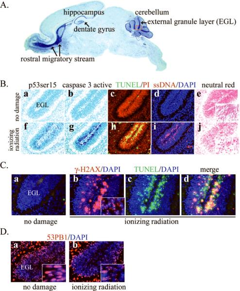
Figure 1. Immunohistochemical analysis of apoptosis after DNA damage in the postnatal mouse brain.
(A) Susceptible areas to DNA damage-induced apoptosis during postnatal brain development include the rostral migratory system, the hippocampus including the dentate gyrus, and the cerebellum particularly the external granular layer (EGL). A sagittal section of a postnatal day 7 mouse brain with Nissl staining is shown. The red box indicates an example of the cerebellar EGL shown in B–D.
(B) Immunohistochemical detection of apoptosis process in the EGL of postnatal day 5 brain induced by ionizing radiation. The lower panel shows activation of p53 (f) and caspase 3 (g), TUNEL positive signal (h), single strand DNA immunopositive signal (i), and pyknosis visualized by Neutral Red staining (j) induced by DNA damage. Inset panels in e and j are magnified views to illustrate the morphology of pyknotic cells. Apoptotic cells are not evident without any insult to the developing brain (a–e). p53 and caspase 3 immunoreactivity were visualized with the VIP substrate kit and the brains were counterstained with Methyl Green (a–b, f–g). TUNEL was done using ApopTag Fluorescein in situ Apoptosis Detection Kit with DAPI counterstaining (c, h). ssDNA antibody was detected with Cy3 conjugated secondary antibody with PI counterstaining (d, i).
(C) Phosphorylated H2AX (γH2AX) is a measure of DNA double strand breaks, and are visualized as nuclear punctate staining (inset panel in b), called foci. In the absence of DNA damage no γH2AX signal is found (a). γ-H2AX staining is detected after DNA damage using Cy3 secondary antibodies (b) and apoptosis is detected by TUNEL using fluorescein (c); panel d is a merge of panels b and c.
(D) Foci formation of 53BP1 after IR induced DNA breaks. Similar to γH2AX, 53BP1 is another early responder to DNA double strand breaks. While 53BP1 is distributed evenly in the nucleus without any DNA damage, 53BP1 foci form in the nucleus after ionizing radiation (b) and were visualized using Cy3-coupled secondary antibodies.

