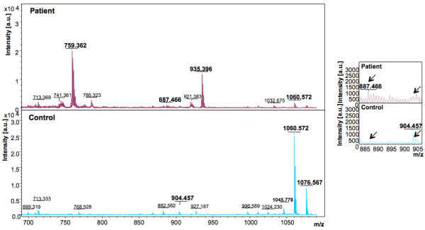
Figure 3.

Representative mass spectra showing peaks in the Reflectron Positive (RP) mode. The upper and lower panels show peak patterns for representative plasma samples from HEV patients and healthy controls, respectively. Peaks in the m/z range of 700 to 1100 are shown. The upper and lower panels show peak patterns for representative plasma samples from HEV patients and healthy controls, respectively. Averaged mass spectra were generated by ClinPro Tools software (V2.0) using spectra showing the highest number of peaks and signal-to-noise ratios. All spectra were baseline-subtracted, smoothed, normalised and realigned. Insets show discriminating peaks not readily apparent at the scale of the main figure.
