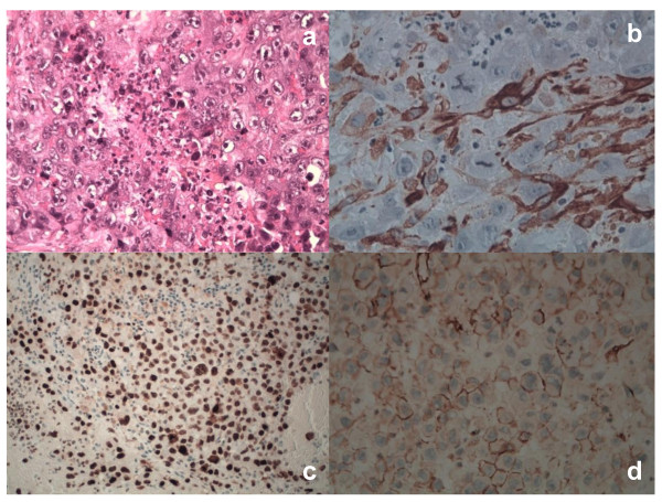
Figure 4.
Histological and immunohistochemical profile of PECs. Tumor cells showed marked coagulative necrosis (Hematoxylin-Eosin, x40) (a). In PECs, SMA had a focally cytoplasmic positivity (x40)(b). Nuclear MIB-1 reactivity was documented in PECs (x200)(c). We observed a strong and diffuse membrane reactivity for CD31 in tumor cells (x40)(d). The tumor was negative for all other markers mentioned, including S-100, CKAE1/AE3, CK5, CD30.

