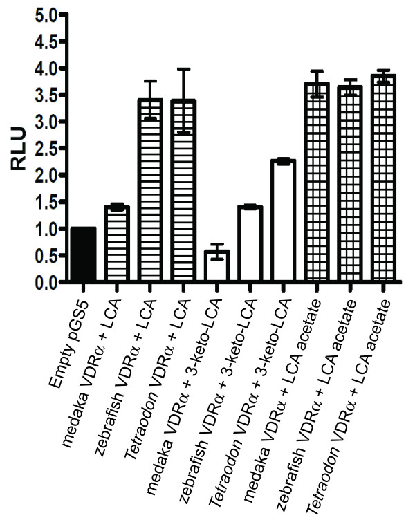
Figure 5.
Transactivation of full-length teleost VDRs. HepG2 cells were transiently transfected with pRL-CMV, XREM-Luc and either medaka VDRα-pSG5, zebrafish VDRα-pSG5, or Tetraodon VDRα-pSG5 as described in Methods. Cells were exposed to 100 μM of either lithocholic acid (LCA), 3-keto-LCA, or LCA acetate for 24 hours. VDR response was measured via dual-luciferase assays. Data is represented as the mean fold induction normalized to control (DMSO) ± SEM.

