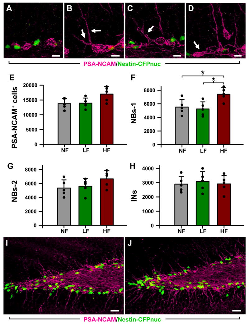
Figure 5. High frequency stimulation of the ATN induces changes in the number of neuroblasts.
A: Type-1 neuroblasts (NB1s), located along the SGZ, with small round soma and short thin horizontal processes. B,C: Type-2 neuroblasts (NB2s) cells located along the SGZ and with horizontal and oblique processes entering the GCL (arrows).
D: Immature neurons (INs), located in the SGZ and the GCL, with a single apical process crossing the GCL. An NB1 cell can also be observed (arrow).
E-H: The overall number of neuroblasts (PSA-CAM+ cells) is slightly higher in the HF animals. Within that group, the number of NB1s is significantly increased (F), whereas the number of NB2s (G) and INs (H) is not changed. I and J Representative confocal microscopy images of sections from the DG of LF (I) and HF (J) mice showing the location of the neuroblasts (PSA-NCAM+ cells, in magenta) and neuroprogenitors (CFPnuc+ cells, in green). Both types of cells are in close apposition. Data are shown as mean ± s.e.m (n = 5 mice for the NF group, and 6 for the LF and HF groups, animals represented by black dots). * p < 0.05. Scale bar is 5 μm in (A-D) and 25 μm in (I, J).

