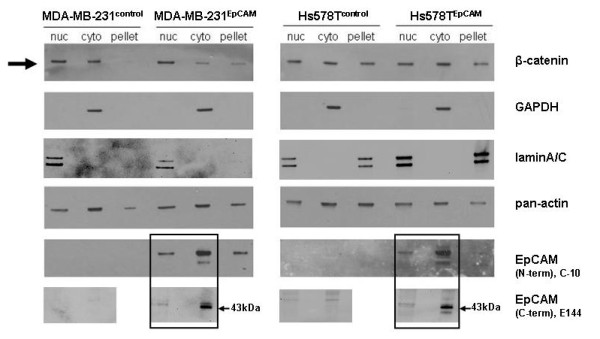
Figure 8.

Subcellular fractionation analysis. In Hs578TEpCAM and MDA-MB-231EpCAM cells and their respective control cell lines, β-catenin was highly expressed in all cell lines, but only in the MDA-MB-231EpCAM cell line a significant and reproducible accumulation of β-catenin could be observed in the nuclear compartment, which resulted in an increased Wnt signaling activity. The mainly cytosolic protein GAPDH was used to control the purity of the cytosolic fractionation. To show integrity of the nuclear fraction lamin a/c antibody was used. Pan-actin was used as loading control. As cells were cultivated to 70-80% density, a majority of the EpCAM protein was present in the cytosol. Additionally to the N-terminal directed anti-EpCAM antibody C-10, EpCAM was detected with a C-terminal directed antibody (E144). Interestingly, EpCAM could be detected in the nuclear fraction with both antibodies.
