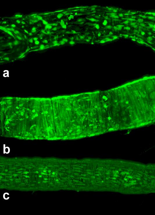
Figure 7.
Examples of effective adenoviral/GFP transfection of lymphatic endothelial and muscle cells in isolated rat mesenteric lymphatic vessels. Vessel diameters are ~ 120-140 μm. Average 2-D projections of the stacks of confocal images, taken at steps of 0.5 μm through the vessels pressurized at 5 cm H2O in the standard isolated vessel chamber.
a. Transfection of mostly lymphatic endothelial cells was performed using solution containing 1.1*1010 viral particles/ml which was injected only inside of non-denuded vessel; b. Transfection of only lymphatic muscle cells in endothelium-denuded vessels using a concentration of 1.5*1010 viral particles/ml only intraluminally; c. Transfection of both lymphatic endothelial and muscle cells required a solution outside the vessel containing 1.1*1010 viral particles/ml together with an intraluminal solution containing 1.5*1010 viral particles/ml.

