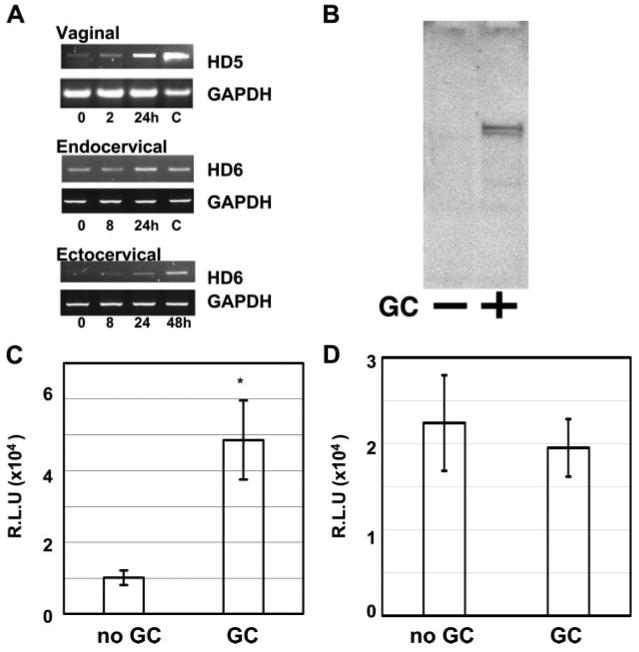
FIGURE 6.

GC infection induces HD5 and HD6 gene expression and enhances HIV infection. A, Immortalized vaginal, endocervical, and ectocervical epithelial cells were infected with N. gonorrhoeae (ATCC no. 43069) at a MOI of 10 and cultured at 37°C. Total RNA was prepared at various time points for RT-PCR analysis. Diluted small intestine cDNA (C, as control; Clontech Laboratories) was included as a positive control for the size of PCR products. The identity of PCR products was confirmed by sequencing. B, Conditioned media from vaginal epithelial cells without (−) or with GC exposure (+) were concentrated and expression of HD5 was analyzed by immunoblotting using anti-HD5 Abs. C, Vaginal epithelial cells were exposed to N. gonorrhoeae at a MOI of 10 and cultured at 37°C for 48 h. Conditioned medium from cells without or with exposure to GC were incubated with pseudotyped HIVJR-FL luciferase reporter virus at 37°C for 1 h before addition to HeLa-CD4-CCR5 cells in the presence of 10% FBS. Difference between conditioned medium from cells without GC exposure and that from cells with GC exposure is significant; *, p < 0.05. Data are means ± SD of triplicate samples and represent two independent experiments. D, To determine the effect of conditioned medium from GC-exposed cells on HIV infection without preincubation with virus, pseudotyped HIVJR-FL luciferase reporter virus mixed with conditioned medium was added to HeLa-CD4-CCR5 cells in the presence of 10% FBS at 37°C for 2 h. Cells were then washed and cultured for 48 h before measurement of luciferase activity. Difference between conditioned medium from cells without GC exposure and that from cells with GC exposure is not significant; p > 0.05. R.L.U., reflective light units.
