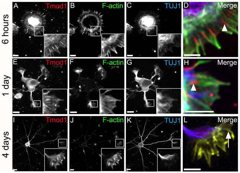
Fig. 2. Tmod1 is associated with F-actin bundles in lamellipodia and growth cones of developing rat hippocampal neurons.
Hippocampal cultures were fixed 6 hours (A-D), 1 day (E-H) or 4 days (I-L) after plating and stained for Tmod1 (A, E and I), F-actin (B, F and J), and β3-tubulin (C, G and K). High magnification insets are displayed as merges (D, H and L). Shown are representative cells from the respective time points. Enrichment of Tmod1 along the proximal part of F-actin bundles in the lamellipodia and filopodia is observed at early times (D, H; arrowheads). Uniform distribution of Tmod1 along F-actin bundles is observed by 4 days after plating (L; arrow). Magnification of insets 2.5× in A-C, 4.3× in E-G and 3.4× in I-K. Scale bars = 10μm except for D, H, and L where scale bars = 5μm.

