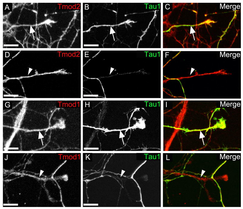
Fig. 3. Tmod2 and Tmod1 are localized to both somatodendritic and axonal compartments in neurons from 7 day-old rat hippocampal neuron cultures.
Hippocampal cultures were fixed 7 days after plating and stained for Tmod2 (A-F) or Tmod1 (G-L). Shown are representative endings of axons as determined by the presence of Tau-1 (A-C and G-I), and dendrites as determined by the lack of Tau-1 (D-F and J-L). Merged images show Tmod (red) and Tau-1 (green) merge (C,F,I and L). Scale bar = 10μm.

