
Figure 5. TNF-α treatment decreases HcrtR2 expression in B35 cells transfected with MSCV-HcrtR2 vector.
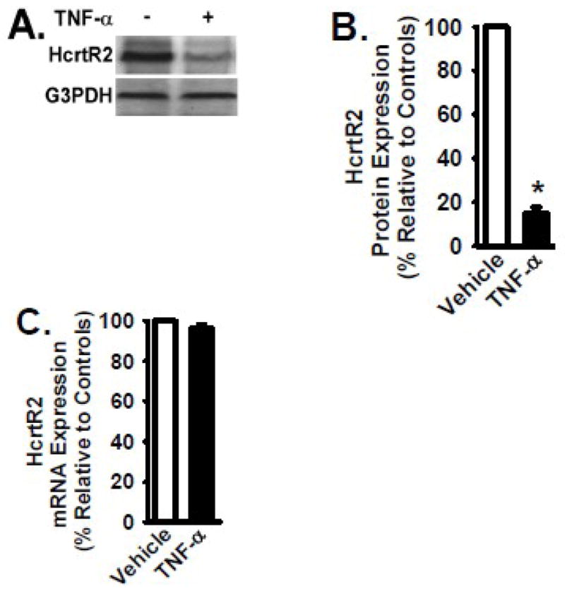
B35 cells overexpressing the HcrtR2 (mediated by MSCV-HcrtR2) were cultured in the presence and absence of TNF-α cytokine (2 ng/ml) for 24 hours at 37°C. Panel A: Cells were lysed, and equivalent amount of whole cell detergent lysates were Western blotted with the indicated antibodies. Panel B: Densitometry of HcrtR2 expression as in Fig. 2. Panel C: mRNA level of HcrtR2 was analyzed by quantitative real time RT-PCR as in Fig. 2. Data are presented as mean ± S.D. (n = 4). * represents p < 0.01 for TNF-α-treated cells compared to vehicle-treated control group.