
Figure 4.
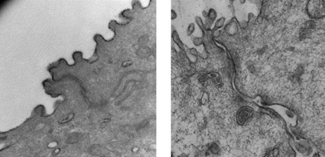
Transmission electron microscopy image (taken at microscope magnification of 53.0K) showing dilated paracellular morphology following a 30 minute time-dose of raised intensity phonation (right panel), compared to control (left panel).
Official websites use .gov
A
.gov website belongs to an official
government organization in the United States.
Secure .gov websites use HTTPS
A lock (
) or https:// means you've safely
connected to the .gov website. Share sensitive
information only on official, secure websites.
Transmission electron microscopy image (taken at microscope magnification of 53.0K) showing dilated paracellular morphology following a 30 minute time-dose of raised intensity phonation (right panel), compared to control (left panel).