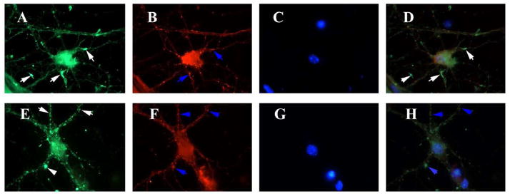
Figure 5. Drp1 distribution relocates from areas of active neurite outgrowth to neuritic processes after amyloid beta treatment.
Hippocampal neurons treated with PBS vehicle or Aβ were immunostained for Drp1, mitochondrial-encoded protein, Cyt. B and nuclear marker, DAPI. Vehicle treated cells (upper panel) showed intense immunoreactivities of Drp1 (A), Cyt. B (B), DAPI (C) and merged (D) in neurite growth cones (white arrows). Drp1 is colocalized with Cyt. B (D) (white arrows). After Aβ treatment (lower panel), Drp1 staining is reduced (E), and Drp1 is colocalzed with mitochondria in merged image (H).

