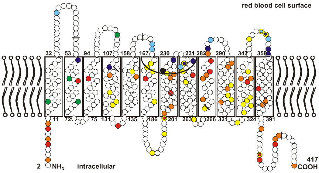
Figure 4. Model of Rhesus proteins in the red blood cell membrane.
Both Rhesus proteins comprise 417 amino acids, shown here as circles. Mature proteins in the membrane lack the first amino acid. The amino acid substitutions that distinguish the RhCE from the RhD protein are marked in yellow, with the 4 amino acids that code for the C antigen in green and the one that codes for the E antigen in black. The single amino acids substitutions which code for partial D are in blue, and those that code for weak D are in red. The mutations that had been identified at the Ulm Institute since 1999 are in light blue and orange. The extracellular Rh vestibule is depicted by the inverted black arc and bordered in part by amino acids of loops 3 and 4. The nine exon boundaries in the RHD cDNA, as reflected in the amino acid sequence, are indicated by black bars.

