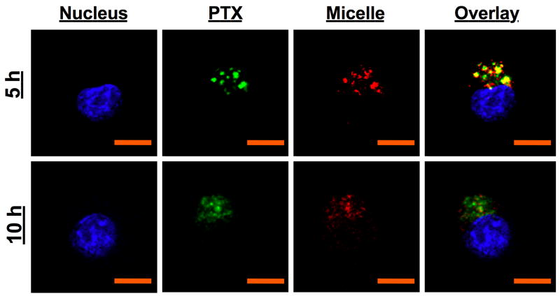
Figure 4.
Confocal microscopy analysis (60X) of the delivery of PTX (AF488, green) encapsulated in folate LDP micelles (AF647, red) in KB cells. Cell nuclei are stained with Hoechst (blue). Scale bar = 10 μm. PTX-folate micelle conjugates are internalized as a whole via FRs and can stay within endosomes for up to 5 h. By 10 h, the low pH of 5.5 and enzymes within the endosome and lysosomes trigger the release of PTX via destabilization of the micelle and breakdown of the polymer, which then diffuses out into the cytosol to cause apoptosis.

