Figure 5.
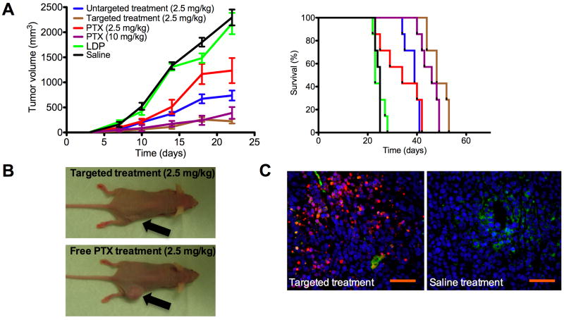
Anti-tumor study in nude mice (n=7) bearing KB xenografts (s.c. injection on right flank, day 0). Treatment began on day 4, when tumors were palpable and mice were given a single dose intravenous injection (tail vein) on day 4, 8, 12 and 16 of the following: (i) untargeted LDP micelles (100 mg/kg) delivering 2.5 mg/kg PX, (ii) folate targeted LDP micelles (100 mg/kg) delivering 2.5 mg/kg PTX, (iii) 2.5 mg/kg free PTX in 1:1 Cremophor:EtOH (1 %v/v) as excipient, (iv) 10 mg/kg free PTX in 1:1 Cremophor:EtOH (1 %v/v) as excipient (v) LDP micelles (100 mg/kg) without PTX and (vi) Saline. Mice were evaluated over a period of 60 days. A) Tumor volume over the first 22 days of treatment. Data is given in mean±SEM for n=7. Kaplan-Meier plot of survival times for mice receiving treatments also show that the targeted therapy had the best long-term efficacy. B) Representative mouse at day 20 (2 days after last treatment) for folate targeted and free PTX treatments at the same dosage level of 2.5 mg/kg. C) Representative histological sections of tumors extracted from mice receiving targeted (ii) and control (vi) treatment. Cell nuclei = blue (Hoechst); Blood vessels = green (CD31-FITC); Apoptotic cells = red (TUNEL, A647). Presence of apoptotic cells far away from blood vessels suggests that micelles were able to penetrate tumor tissue effectively. Scale bar = 250 μm. (Data in mean ± SEM)

