
Figure 3.
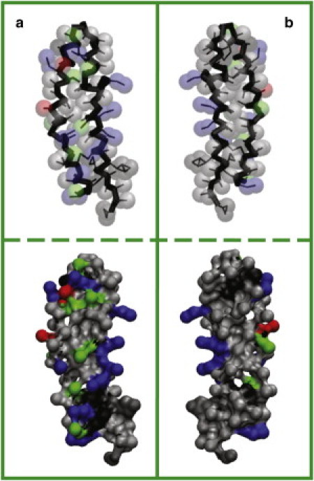
Distribution of residues of (a) water- and (b) membrane-facing sides of SP-B. In the upper panel, bonds are shown as thin black lines, backbone as thick black lines, and side chains as semitransparent spheres. Apolar residues are colored in gray, polar in green, positively charged in blue, and negatively charged in red. The solvent-accessible surface with the same color scheme is given in the lower panel.