
Figure 5.
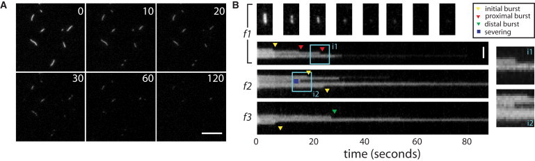
Single actin filaments disassemble in bursts in Xenopus egg cytoplasmic extracts. (A) Timelapse images of single actin filaments perfused with Xenopus egg extract. Elapsed time in seconds is shown in upper right of each image. Scale bar = 10 μm. (B) Kymographs of representative filaments (f1–f3), showing time on the x axis and distance along the filament contour on the y axis. (Triangles) Events scored as endwise bursting; (yellow triangles) initial bursts. (Red and green triangles) Proximal and distal bursts, respectively. (Blue squares) Internal events scored as severing events. (Light-blue square) Regions i1 and i2 magnified (insets on right). Filament polarity is unknown in these experiments. Scale bar = 2 μm.