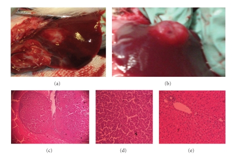
Figure 2.
Macroscopic and microscopic morphology of secondary liver tumors. VX2 tumor cell grafts were implanted in liver from rabbits 14 days prior to tumor removal. All implanted tumor grafts showed macroscopic growth on liver sites. Some growths were observed to have a flat growth (a) while some others have a nodular growth (b). At histology, most tumors had a pseudocapsule formed by fibrotic tissue surrounded the tumor ((c) x20). Typical epithelial cells with malignant morphology are seen, however no neoangiogenesis, tumor invasion, or lymphocyte infiltration was seen at this stage of tumor growth ((d) x40). Typical normal liver rabbit tissue is seen in the rest of the organ ((e) x40).

