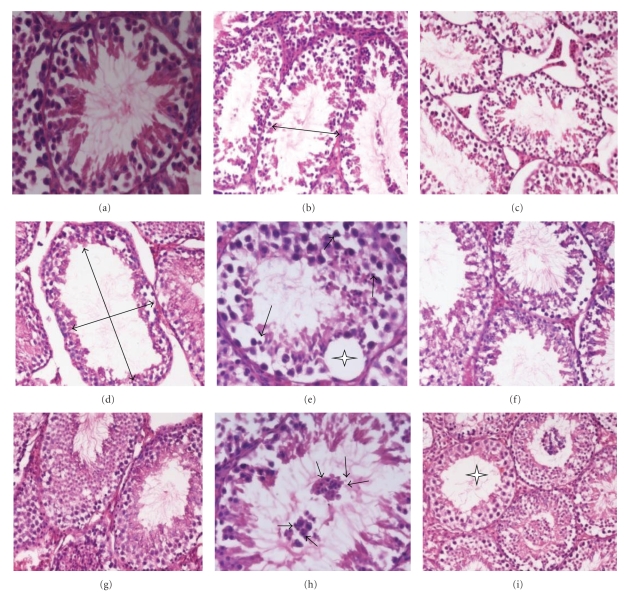
Figure 1.
Modulation of radiation-induced histological changes in testes of Swiss albino mice by T. cordifolia extract. Photomicrograph (a) and (b) showing normal architecture of testis of sham irradiated and negative control, respectively. Photomicrographs (b, c, d, e) are showing radiation-induced pathological changes in irradiated control group from days 3, 7, 15, and 30, respectively, in the form of disrupted germinal epithelium, empty tubule (large arrow), intertubular oedema, pycnotic nuclei (small arrow), and intraepithelium vacuolation (asterick) with depleted germ cells population. Photomicrographs (f, g, h, and i) are showing better testicular architecture in TCE-treated irradiated group from days 3, 7, 15, 30, respectively, with a multinucleated formation of round spermatids (small arrow) at day 15 and almost normal structure at day 30 of experiment.

