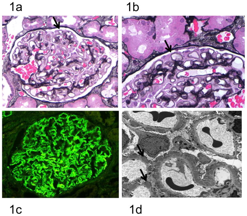
Figure 1. Human Membranous Nephropathy Pathology.

At early stages, the glomeruli and interstitium look essentially normal, but as the disease progresses, the thickening of capillary loops becomes evident due to the accumulation of sub-epithelial immune complexes and the deposition of new basement membrane material by the podocyte (Figure 1a, ×400). Staining with silver methenamine may reveal characteristic spikes (arrowed) representing basement membrane material projecting between the immune deposits (Figure 1b, ×1000). Immunofluorescence reveals finely granular deposits of IgG (predominantly IgG4) in a subepithelial distribution on the outer surface of the GBM (Figure 1c). Electron microscopy reveals the characteristic subepithelial immune deposits (arrowed) (Figure 1d).
