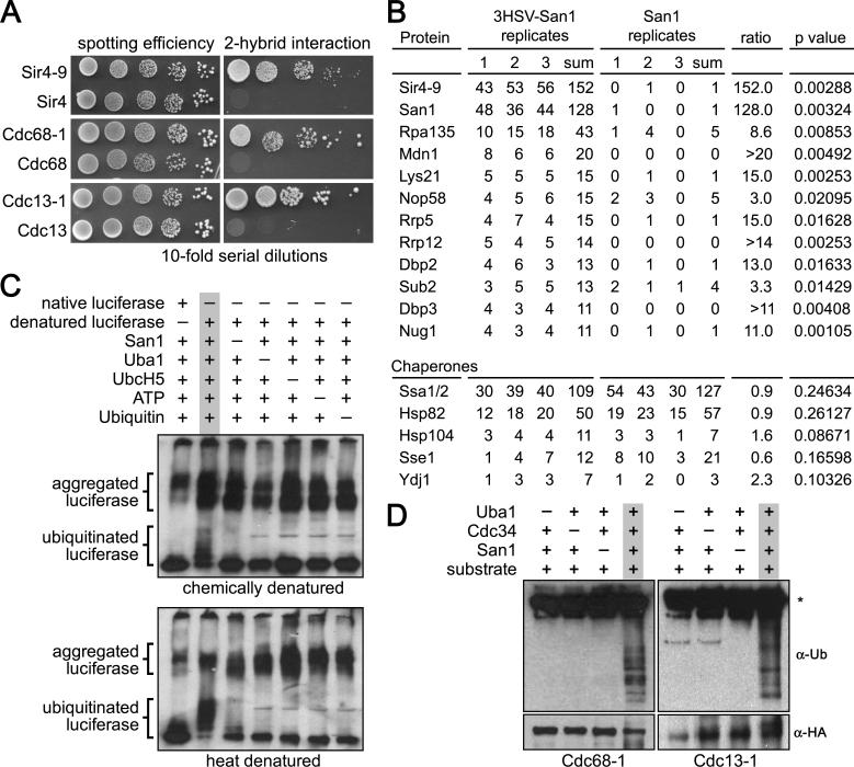
Figure 1. San1 directly interacts with substrates.
(A) Cells expressing GBD-San1C279S and each GAD fusion were spotted onto media plus or minus histidine to measure spotting efficiency and 2-hybrid interaction. (B) The top 12 proteins with ≥3-fold enrichment in the 3HSV-tagged coIP are shown at the top. Chaperones known to function in PQC degradation are shown at the bottom. Numbers are spectral counts for each protein in each replicate (C) Chemical or heat denatured luciferase was added to a reaction containing the San1 ubiquitination cascade. Western blot was probed with anti-luciferase antibody. Dashes above the lanes indicate which reagent was excluded from the reaction. (D) 2xHA-tagged substrates were immunoprecipitated from E. coli cells expressing the San1 ubiquitination cascade. Western blots were probed with anti-ubiquitin antibodies to assess substrate ubiquitination or anti-HA antibodies to assess substrate immunoprecipitation. Dashes above the lanes indicate which components were not expressed. Asterisk marks the antibody band.

