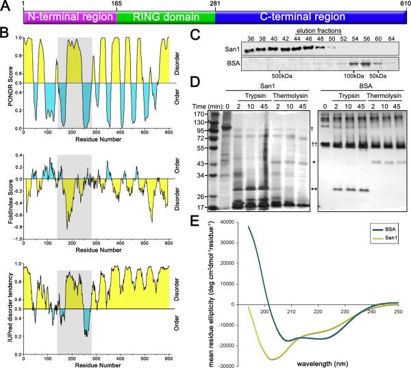
Figure 2. San1 is disordered.
(A) A cartoon representing overall topology of San1. (B) Disorder predictions of San1. Top panel is PONDR (http://www.pondr.com/). Middle panel is FoldIndex (http://bip.weizmann.ac.il/fldbin/findex). Bottom panel is IUPred (http://iupred.enzim.hu/index.html). Predicted disordered regions are in yellow and predicted ordered regions are in blue. San1's RING domain is in gray. (C) Purified San1 or BSA was loaded onto a Superdex 200 column and eluted with 50mM NaCl, 15mM Na2PO4, pH7.3. Western blot of San1 was probed with anti-HSV antibodies. BSA gel was stained with Coomassie. (D) Purified San1 or BSA was incubated with trypsin or thermolysin for the indicated times. Proteins were separated by SDS-PAGE and visualized by silver stain. The locations of trypsin (*), thermolysin (**), San1 (†), and BSA (††) are marked. (E) CD spectra of San1 (yellow) and BSA (blue) were recorded at 0.2 mg/ml for San1 and 0.15 mg/ml for BSA in 50mM NaCl and 15mM Na2HPO4, pH7.3 at 25°C.

