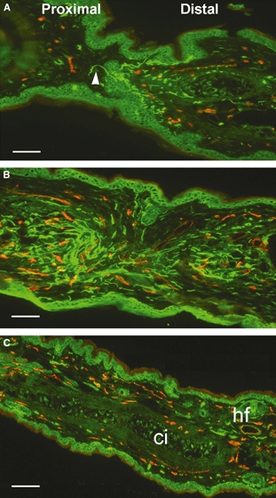
Fig. 5.

Reinnervation and vascularisation of MRL/MpJ ear at 84 days post-wounding. Sequential sections through the centre of an ear wound (A) show fusion of opposing epithelial margins and penetration of pioneer regenerating nerve fibres (arrowhead) from the proximal into distal blastema. (B) Disintegration of dividing epithelium and further infiltration of regenerating nerves from the proximal wound margin accompanied by new blood vessels. Away from the centre of the wound where tissue regeneration was nearly complete (C) evident from fusion of cartilage islands (ci) and emergence of hair follicles (hf), nerve and blood vessel density notably decreased, resembling that of an unwounded mouse ear. Scale bars = 100 μm.
