
Fig. 6.
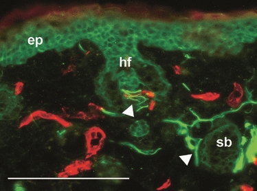
Innervation of hair follicles in newly regenerated MRL/MpJ ear tissue. Dual immunohistochemical staining for pan-neurofilament (fluorescein isothiocyanate, green) and CD31 (tetramethyl rhodamine isothiocyanate, red) of the MRL/MpJ mouse ear at 84 days post-wounding. Autofluorescence shows structures such as epidermis (ep), hair follicles (hf) and sebaceous glands (sb). Arrowheads show innervation of target structures within the newly regenerated tissue of the MRL/MpJ ear wound. Scale bar = 100 μm.