
Fig. 8.
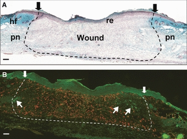
Innervation and vascularisation of MRL/MpJ dorsal skin excisional wounds. Masson's trichrome-stained section of a day 7 full-thickness dorsal skin wound (A) depicts wound area (arrows and dotted line), re-epithelialisation of wound (re), excised panniculus (pn) and surrounding hair follicles (hf). Immunohistochemical dual staining for pan-neurofilament (fluorescein isothiocyanate, green) and CD31 (tetramethyl rhodamine isothiocyanate, red) was used to detect regeneration of nerves and blood vessels into the wound. At 7 days post-wounding (B), blood vessels were the first to form in the wound area followed by only a few nerve fibres from the periphery (arrows). Scale bars = 100 μm.