
Fig. 2.
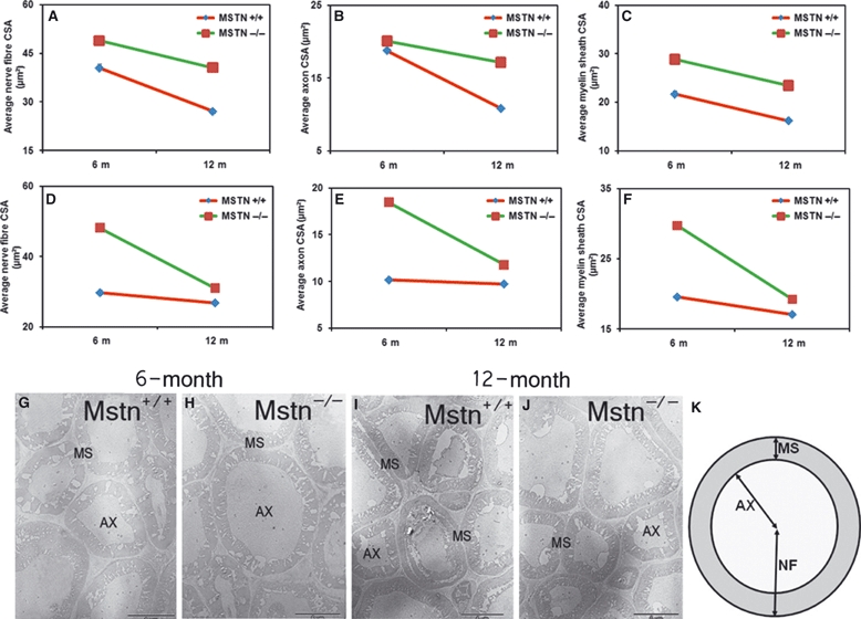
Myostatin deletion increases nerve fibre CSA. Average nerve fibre CSA (axons and myelin sheath) of the radial (A) and ischiatic (D) nerves of 6- and 12-month-old mstn+/+ and mstn−/− mice. (A) mstn−/− mice show a significant increase in nerve fibre CSA compared to mstn+/+ counterparts (P < 0.001). Significant age-related changes in CSA were observed in both genotypes analysed (P < 0.001). Average axon CSA of the radial (B) and ischiatic (E) nerves of 6- and 12-month-old mstn+/+ and mstn−/− mice. Two-way anova for both nerves showed significant changes by genotype, age and interaction of the two parameters. Average myelin sheath CSA of the radial (C) and ischiatic (F) nerves of 6- and 12-month-old mstn+/+ and mstn−/− mice. Two-way anova for both nerves showed significant changes by genotype, age and interaction of the two parameters. (G–J) TEM images showing sections of radial nerve from 6- and 12-month-old mstn+/+ and mstn−/− mice. Images from 6-month-old mstn−/− mice show an increase in the CSA of axons compared to age-matched mstn+/+ animals. Fibres from 12-month-old mstn−/− radial nerves show a reduction in the CSA compared to those from 6-month-old animals. AX, axon; MS, myelin sheath. (K) Diagrammatic illustration showing the technique used for nerve fibre (NF), axon (AX) and myelin sheath (MS) cross-sectional area measurements. Scale bar: 5 μm.