
Fig. 5.
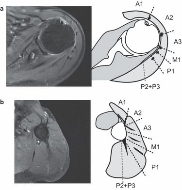
Dividing method of the deltoid muscle on MR images. The intramuscular tendons are clearly depicted in a T2-weighted transverse MR image with fat suppression. The deltoid muscle is divided based on the distribution of intramuscular tendons. The straight lines show the border between each muscle segment. The proximal intramuscular tendons locate at the borders of each segment. For the distal insertions, A3, M1 and P1 intramuscular tendons exist at their center. On the other hand, A1, A2 and P3 have their distal intramuscular tendons at the margin of the segments. (a) At proximal deltoid level, (b) At distal deltoid level.