
Fig. 1.
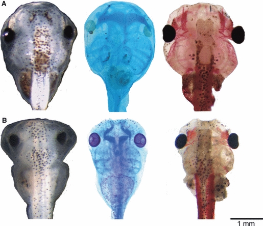
Effects of FoxN3-MO injections on the cranial external morphology, cartilage anatomy (Alcian-stained cartilage appear dark blue), and muscle morphology (muscle stained with newt skeletal muscle antibody) in X. laevis larva at Stage 46. (A) Co-MO-injected tadpole with normally formed skull (left), cartilage (middle), and muscle (right) anatomy. (B) FoxN3-MO (bilaterally)-injected tadpole has a significantly smaller skull with several oedema formations in head and trunk region (left), combined with smaller and malformed cartilage (middle) and muscle (right) results from FoxN3-MO injection. All images are dorsal views of larval skulls with the anterior up.